Paper explores physical mechanisms of particle separation in viscoelastic fluids and its optimization
block Heading link
A joint research paper published by Zhangli Peng, an assistant professor and member of the Center for Bioinformatics and Quantitative Biology, and Ian Papautsky, a Richard and Loan Hill Professor, in the Department of Biomedical Engineering, and their teams studied the effects of flowrate, the speed at which fluid moves, particle size, and the shear-thinning extent of the fluid on separating and focusing particles in viscoelastic fluids.
This team wanted to simulate design scenarios focused on studying physical mechanisms and optimizing separation. They specifically wanted to focus on how to dig into the physics of how separation happens, as it is not quite clear. This work can help them to fine tune the design of devices.
“We used different methods to simulate the same behavior and once we simulate those processes, we can talk more about the underlying physical mechanism in separating particles,” said Moein Naderi, a graduate student who is first author of the paper. “We also have a better understanding of and can fabricate devices without running many experiments.”
Viscoelastic fluids are non-Newtonian fluids, including biologically related fluids such as blood or saliva, that behave differently than water, a Newtonian fluid.
“We think if we work with viscoelastic flow, it might be easier to process bio-fluid samples directly, which would save costs while detecting and diagnosing diseases,” Peng said.
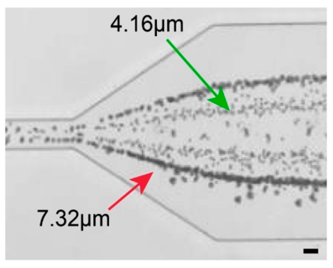
Expansion region the channel outlet enabling separation of the focused streams. (Scale bar in all images: 25 μm).
This research of separating bioparticles has the potential to catch some bacteria and diseases early on, such as sepsis, a very small but potentially deadly bacteria, able to harbor in the blood stream that needs to be detected early to save a patients’ life.
Peng’s modeling lab collaborated with Papautsky’s experimental lab to utilize the computational modeling simulations, as well as the experimental aspect of microfluidic technologies.
“Whereas many groups usually conduct only experiments or simulations, we combined both to complement each other,” Peng said.
The paper, “Elasto-Inertial Focusing Mechanisms of Particles in Shear-Thinning Viscoelastic Fluid in Rectangular Microchannels,” in the Micromachines journal was written by Peng and Papautsky and their team including Jian Zhou, a research assistant professor, and graduate students, Naderi and Ludovica Barilla, a graduate student in UIC’s dual master’s degree program with the Politecnico di Milano in Italy, who did the microfluidic experiments.
They are also working on an additional paper that focuses on showing the impact of cell shapes and how those can be used to uphold the separation and applications within microfluidics.